March 19, 2012 weblog
Ticks found able to survive being subjected to electron microscopy

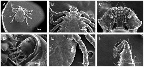
(PhysOrg.com) -- Most people know that ticks are rather hardy little creatures, killing them generally takes some severe bashing with a blunt object, or incineration in an open fire. But few likely suspected they would be able to withstand the extreme conditions a typical specimen undergoes in order to be viewed properly under a scanning electron microscope (SEM). But, clearly they can as Yasuhito Ishigaki and his colleagues at Japan’s Kanazawa Medical University recently discovered. As they describe in their paper published in PloS One, most of the ticks they placed in their SEM survived not only the ordeal, but were able to walk away afterwards.
A SEM works by recording how electrons are scattered or absorbed when fired at a specimen, which is of course rather harsh treatment. Making things even harsher is the fact that in order to create the very sharpest images possible, the specimen is held in a vacuum to prevent air and particles from messing up the image. Most organisms would succumb in minutes to either condition, but somehow, the ticks managed to survive and to be taped doing so. In the video the team made, the ticks’ legs can be seen moving. And if that weren’t enough, the ticks were able to walk away once they were removed from the SEM, proof positive that ticks will now have to be added to the list of hardiest organisms on the planet.
The ticks didn’t go unscathed however, as they all died within a couple of days after being scanned, rather than live another couple of weeks which would have been the norm. Also, in true horror-movie fashion, the ticks, eerily, seemed to be trying to fold their legs in during the electron blast to avoid having them cooked.
The team reports that they stumbled into their experiment with the ticks and SEM when Ishigaki found some ticks had survived being subjected to a vacuum chamber involved in a different procedure. They also report that they’re almost certain the shortened life span brought about by being scanned by the SEM was due to the electron beam blast rather than the vacuum because they subjected some of the ticks to just the vacuum and found they lived as long as they normally would have afterwards.
More information: Ishigaki Y, Nakamura Y, Oikawa Y, Yano Y, Kuwabata S, et al. (2012) Observation of Live Ticks (Haemaphysalis flava) by Scanning Electron Microscopy under High Vacuum Pressure. PLoS ONE 7(3): e32676. doi:10.1371/journal.pone.0032676
Abstract
Scanning electron microscopes (SEM), which image sample surfaces by scanning with an electron beam, are widely used for steric observations of resting samples in basic and applied biology. Various conventional methods exist for SEM sample preparation. However, conventional SEM is not a good tool to observe living organisms because of the associated exposure to high vacuum pressure and electron beam radiation. Here we attempted SEM observations of live ticks. During 1.5×10^−3 Pa vacuum pressure and electron beam irradiation with accelerated voltages (2–5 kV), many ticks remained alive and moved their legs. After 30-min observation, we removed the ticks from the SEM stage; they could walk actively under atmospheric pressure. When we tested 20 ticks (8 female adults and 12 nymphs), they survived for two days after SEM observation. These results indicate the resistance of ticks against SEM observation. Our second survival test showed that the electron beam, not vacuum conditions, results in tick death. Moreover, we describe the reaction of their legs to electron beam exposure. These findings open the new possibility of SEM observation of living organisms and showed the resistance of living ticks to vacuum condition in SEM. These data also indicate, for the first time, the usefulness of tick as a model system for biology under extreme condition.
via DiscoverBlogs
© 2011 PhysOrg.com


