December 24, 2019 feature
MATRIEX imaging: Simultaneously seeing neurons in action in multiple regions of the brain
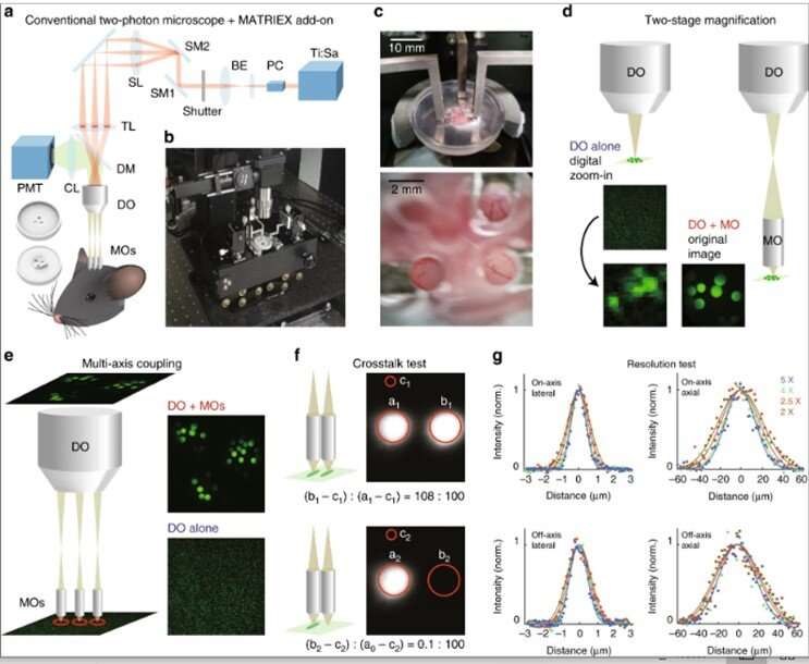
Two-photon laser scanning microscopy imaging is commonly applied to study neuronal activity at cellular and subcellular resolutions in mammalian brains. Such studies are yet confined to a single functional region of the brain. In a recent report, Mengke Yang and colleagues at the Brain Research Instrument Innovation Center, Institute of Neuroscience, Center for Systems Neuroscience and Optical System Advanced Manufacturing Technology in China, Germany and the U.K. developed a new technique named the multiarea two-photon real-time in vitro explorer (MATRIEX). The method allowed the user to target multiple regions of the functional brain with a field of view (FOV) approximating 200 µm in diameter to perform two-photon Ca2+ imaging with single-cell resolution simultaneously across all regions.
Yang et al. conducted real-time functional imaging of single-neuron activities in the primary visual cortex, primary motor cortex and hippocampal CA1 region during anesthetized and awake states in mice. The MATRIEX technique can uniquely configure multiple microscopic FOVs using a single laser scanning device. As a result, the technique can be implemented as an add-on optical module within existing conventional single-beam-scanning, two-photon microscopes without additional modifications. The MATRIEX can be applied to explore multiarea neuronal activity in vivo for brain-wide neural circuit function with single-cell resolution.
Two-photon laser microscopy originated in the 1990s to become popular among neuroscientists interested in studying neural structures and functions in vivo. A major advantage of two-photon and three-photon imaging for living brains include the optical resolution achieved across densely labelled brain tissues that strongly scatter light, during which optically sectioned image pixels can be scanned and acquired with minimal crosstalk. However, the advantages also caused significant drawbacks to the method by preventing the simultaneous view of two objects within a specific distance. Researchers had previously implemented many strategies to extend the limits, but the methods were difficult to implement in neuroscience research labs. Nevertheless, an increasingly high demand exists in neuroscience to investigate brain-wide neuronal functions with single-cell resolution in vivo.
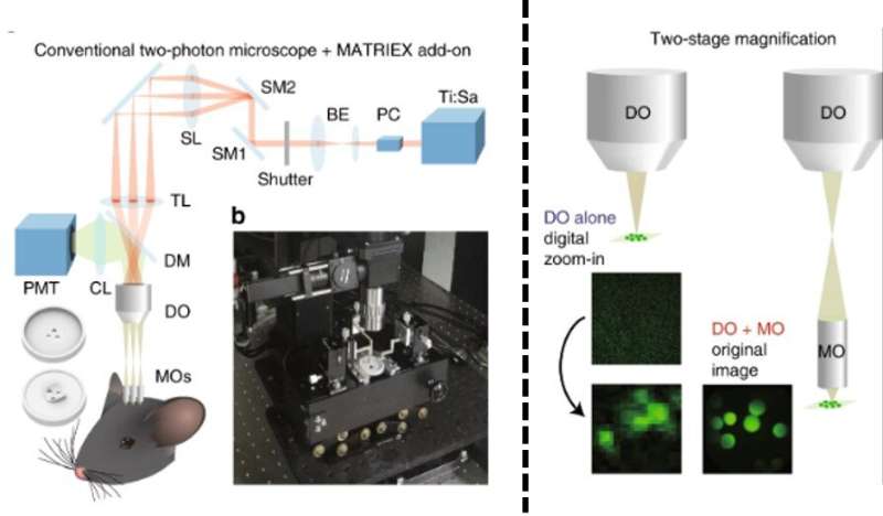
In a straightforward approach, scientists can place two microscopes above the same animal brain to image the cortex and cerebellum simultaneously. But such efforts can lead to substantial increases in complexity and cost. The existing high expectations for performance and feasibility therefore pose a highly challenging engineering question on how a single imaging system can simultaneously obtain live microscopic images from multiple brain regions in vivo. To address the question, Yang et al. introduced a new method that combined two-stage magnification and multi-axis optical coupling.
They realized the method using a low-magnification dry objective (DO), with multiple water-immersed, miniaturized objectives (MOs) under the dry objective. The scientists placed each of the MOs at the desired target position and depth in the brain tissue. The team used the new compound object assembly similarly to the original water-immersed microscope objective without additional modifications to the image scanning and acquisition subsystem.
The research team first assembled the MATRIEX compound objective. For this, they replaced the conventional water-immersion microscope objective with a customized compound objective assembly, inside a two-photon laser scanning microscope equipped with a conventional single-beam raster scanning device. The compound assembly contained multiple MOs (miniaturized objectives) inserted through multiple craniotomies during which the scientists glued a 3-D printed plastic chamber to the skull of the mouse model. The chamber roughly aligned the MOs with the same space to adjust lateral position and depth. Yang et al. precisely manipulated the individual MOs to view the objects under all MOs simultaneously in the same image plane.
They implemented the MATRIEX method using two principles; two-stage magnification and multiaxis coupling. For example, using two-stage magnification with the dry objective (DO) alone, they observed 20 µm beads as tiny blurry dots while observing crisp, round circles through the compound assembly. During multiaxis coupling, the scientists coupled a single DO with multiple MOs on the same image plane. Using a simple raster scan in a single rectangular frame, the research team acquired a rectangular image containing multiple circular FOVs (Field of Views) – where each FOV corresponded to one MO with minimal inter-FOV pixel crosstalk.
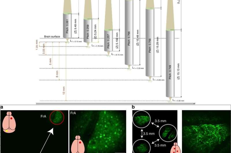
The scientists credited the magnification of the numerical aperture (NA) for allowing better resolution with the compound assembly. The associated lenses were also flexible and custom-designed for mass-production at low cost to assist experimental design. The main feature of MATRIEX was its capacity to image multiple objects simultaneously at large depth intervals. To highlight this, Yang et al. designed different MOs with diverse parameters, placing them at a specific depth where the corresponding object planes conjugated on the same axis. In practice, the research team compensated minor mismatches between the desired and actual object depth by adjusting MOs individually along each of the z axes.
Typically, under the DO (dry objective) the maximum lateral size of the target zone is limited by the maximum size of the scanning field. For example, using a DO with a 2x magnification and target zone of 12 mm in diameter, scientists can image an entire adult mouse brain. In this study, Yang et al. simultaneously imaged the frontal association cortex and cerebellum of the mouse. In practice, a 4x air objective was suited to achieve better resolution to observe fine dendrite structures.
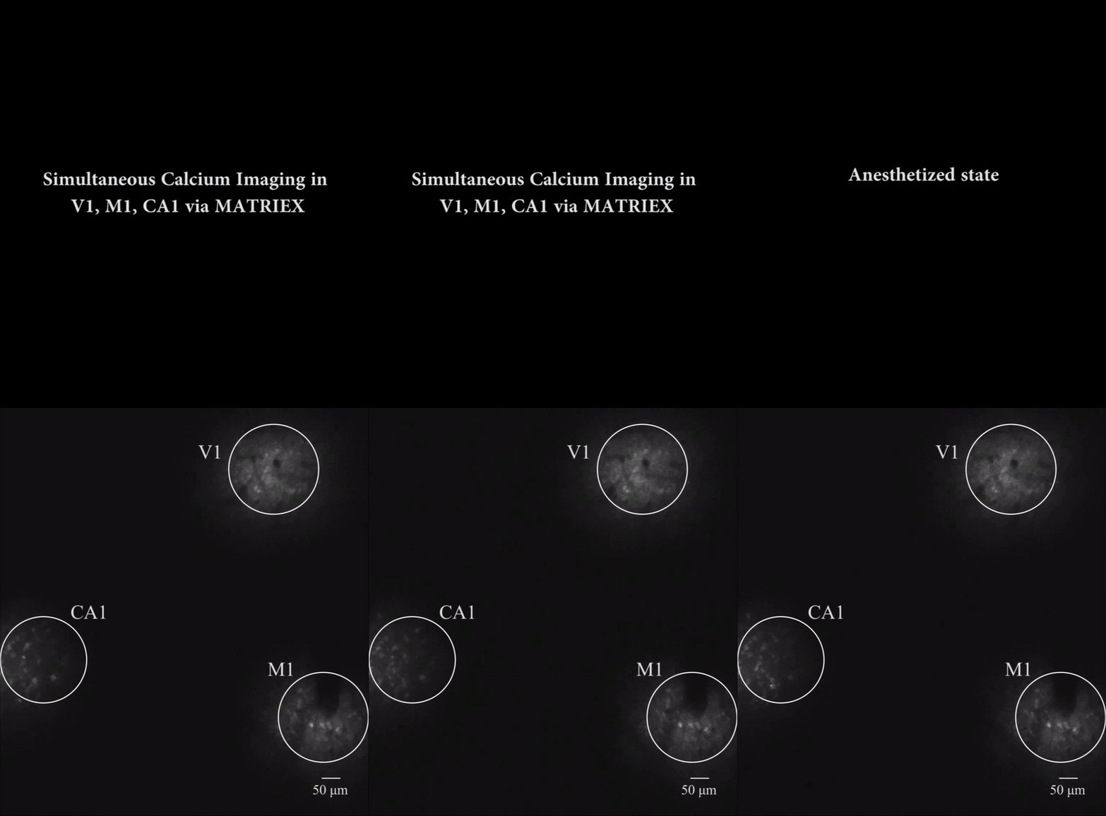
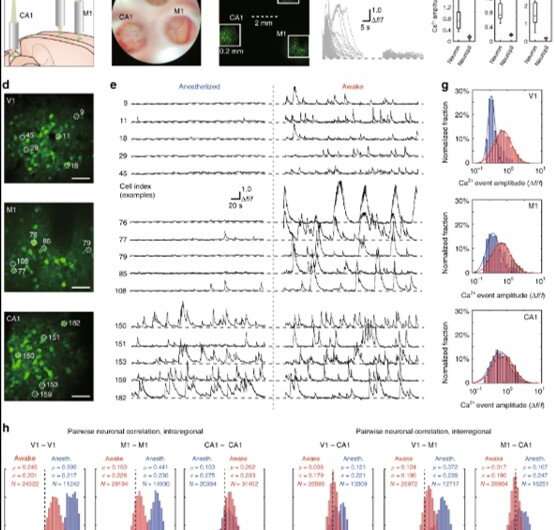
As proof of principle, the research team used MATRIEX to perform simultaneous two-photon Ca2+ imaging of fluorescently-labelled neurons in the primary visual cortex (V1 region), primary motor cortex (M1 region) and hippocampal CA1 region of mice. In the configuration of the three MOs, the scientists placed two MOs suited for the V1 and M1 region, directly above the cortex and inserted an MO within the hippocampal CA1 region after surgically removing a cortical tissue. The team then designed the lenses for the object planes corresponding to V1, M1 and CA1 for conjugation on the same image plane. Using a two-photon microscope equipped with a 12 kHz resonant scanner, the scientists scanned the full image to observe three FOVs and their single cells after enlarging the three different sections to resolve single neurons. Then they noted the laser power to be distributed among multiple FOVs.
While Yang et al. could have obtained these results using conventional single-FOV imaging within a single brain region, the MATRIEX technique provided them data beyond those offered with single-FOV imaging techniques. Taken together, these results allowed a highly inhomogeneous distribution and transformation of spontaneous activity patterns from the anesthetized state to the awake state in mice, spanning a brain-wide circuit level at single-cell resolution.
In this way, Menge Yang and co-workers developed the MATRIEX technique based on the principle of two-stage magnification and multiaxis optical coupling. They simultaneously conducted two-photon Ca2+ imaging in neuronal population activities at different depths in diverse regions (V1, M1 and CA1) in anesthetized and awake mice with single-cell resolution. Importantly, any conventional two-photon microscope can be transformed into a MATRIEX microscope, while preserving all original functionalities. The key to transformation is based on the design of a compound objective assembly. The researchers can use different, carefully designed MOs to suit diverse brain regions with 100 percent compatibility between the MATRIEX technique and conventional microscopy. The research team expect the MATRIEX technique to substantially advance three-dimensional, brain-wide neural circuit dynamics at single-cell resolution.
More information: Mengke Yang et al. MATRIEX imaging: multiarea two-photon real-time in vivo explorer, Light: Science & Applications (2019). DOI: 10.1038/s41377-019-0219-x
Tianyu Wang et al. Three-photon imaging of mouse brain structure and function through the intact skull, Nature Methods (2018). DOI: 10.1038/s41592-018-0115-y
Rongwen Lu et al. Video-rate volumetric functional imaging of the brain at synaptic resolution, Nature Neuroscience (2017). DOI: 10.1038/nn.4516
Journal information: Nature Methods , Light: Science & Applications , Nature Neuroscience
© 2019 Science X Network


