October 22, 2019 feature
Extreme biomimetics – the search for natural sources of materials engineering inspiration
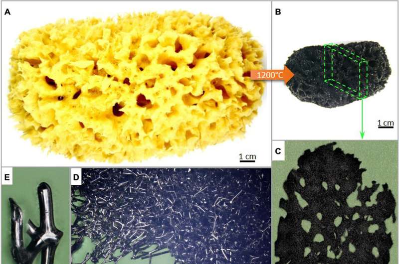
Biologically inspired engineering to produce biomimetic materials and scaffolds typically occurs at the micro- or nanoscale. In a new study on Science Advances, Iaroslav Petrenko and a multidisciplinary global research team, proposed the use of naturally pre-fabricated, three-dimensional (3-D) spongin scaffolds to preserve molecular detail across larger, centimeter-scale samples. During materials characterization studies, researchers require large-scale samples to test nanoscale features. The naturally occurring collagenous resource contained a fine-scale structure, stable at temperatures of up to 12000C with potential to produce up to 4 x 10 cm 3-D microfibrous and nanoporous graphite for characterization and catalytic applications. The new findings showed exceptionally preserved nanostructural features of triple-helix collagen in the turbostratic (misaligned) graphite. The carbonized sponge resembled the shape and unique microarchitecture of the original spongin scaffold. The researchers then copper electroplated the composites to form a hybrid material with excellent catalytic performance observed in both fresh water and marine environments.
Extreme biomimetics is the search for natural sources of engineering inspiration, to offer solutions to existing synthetic strategies. Bioengineers and materials scientists aim to create inorganic-organic hybrid materials that are resistant to harsh chemical and thermal microenvironments to mimic naturally prefabricated 3-D architecture. For example, scientists have used marine sponges as a productive model system to develop new, hierarchically structured 3-D composites with renewable, non-toxic organic scaffolds. During its evolution 600 million years ago, marine demosponges had produced constructs ranging from the centimeter to meter scale, with potential applications at present in materials research.
The fibrous component of the sponge skeleton known as spongin, belongs to the collagen suprafamily and is the focus in materials engineering due to its nano-architectural organization and biomechanical behavior. Structurally, collagen-like spongin has multiple levels, consisting of 100 µm-thick single fibers and nanofibers, combined into complex 3-D hierarchical networks of high macro-porosity. Due to spongin's thermostability of up to 3600C and its resistance to acids, researchers have used spongin-based scaffolds in hydrothermal synthesis reactions to develop ferrous oxide (Fe2O3) and titanium dioxide (TiO2-)-based composites for electrochemical and catalytic purposes. Scientists had also carbonized spongin-scaffolds to develop centimeter-scale manganese dioxide (MnO2)-based supercapacitors.
In current trends in materials science, scientists aim to develop carbon materials with controlled microarchitectures and morphologies at large scales using renewable and biodegradable natural sources. Recent studies have recommended the suitability of structural proteins such as keratin, collagen and silk for carbonization between 2000C to 8000C and even up to 28000C in temperature. Nevertheless, studies on sponge-like, ready-to-use carbon scaffolds with hierarchical pores and 3-D connected skeletons hitherto remain unreported.
As a result, Petrenko et al. developed new 3-D carbonized spongin scaffolds by combining hierarchical complexity from the nanometer to centimeter scale, capable of withstanding temperatures greater than 12000C, while retaining nanoscale architecture. The research team hypothesized the possibility of converting spongin to carbon at high temperatures, without loss of its form or structural integrity to favor its functionalization into a catalyst. In the new work, they detailed the first successful effort to design a centimeter-scale 3-D carbonized spongin Cu/Cu2O catalytic material using an extreme biomimetics strategy. The research team then demonstrated the ability of the material to effectively catalyze the reduction of 4-nitrophenol (4-NP) to 4-aminophenol (4-AP) in fresh water and marine environments.
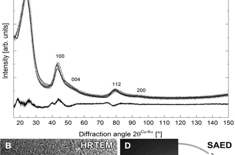
The scientists first heated the sponge skeletons to directly carbonize them. The carbonized spongin decreased in volume but maintained a 3-D fibrous appearance and an increased density compared to native spongin. The research team then analyzed the carbonaceous material using 13C nuclear magnetic resonance (NMR) spectroscopy to understand its structural chemistry. Compared to previous results, the team found the material to resemble amorphous graphite containing ordered, graphite-like domains. They confirmed the findings using X-ray diffraction (XRD) and Raman spectroscopy. The team confirmed the constitution of the graphite (obtained from spongin) using high-resolution transmission electron microscopy (HRTEM), fast Fourier transformation (FFT) and selected-area electron diffraction (SAED) techniques. The electron energy-loss spectroscopy spectra (EELS) measurements for carbonized spongin corresponded with previous results.
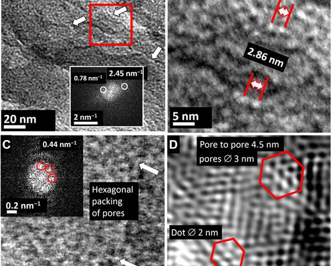
At the nanoscale, the graphite nanoclusters produced a porous structure, which Petrenko et al. investigated using a TEM (transmission electron microscopy) micrograph of the carbonized sponge to reveal a collagen-based fibrillar protein. They observed nanostructures with pearl-like chains and periodicities, as well as the preservation of structural features of the collagen helix after carbonization of spongin. Fourier transform images revealed a hexagonal lattice at the nanoscale and the scientists verified the transformation of collagen-based spongin into a hexagonal carbon structure. The research team then systematically investigated the structural and chemical changes of carbonization using additional materials characterization techniques. The results showed the gradual evolution of the material from carbon toward nanocrystalline graphite.
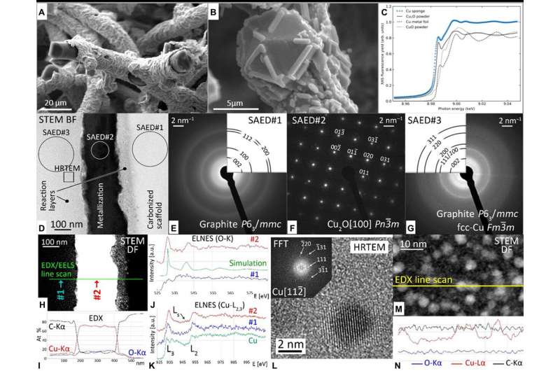
Since the electrical conductivity of carbon is a well-recognized property, the team functionalized the carbonized spongin scaffolds with copper using the electroplating method. After Petrenko et al. electroplated the material sample with copper (Cu) for 30s, the resulting 3-D carbonized scaffold resembled the architecture of the material prior to metallization. They then used Raman spectroscopy, XPS and X-ray absorption spectroscopy to identify the phases of Cu within the Cu/Cu2O carbonized spongin scaffolds (known as CuCSBC). They followed the investigations using chemical and structural studies of the new, catalytic CuCSBC material.
The research team then tested the reduction reaction of 4-nitrophenol (4-NP) to 4-amino phenol (4-AP) in the presence of CuCSBC. Typically, 4-NP constitutes pharmaceutical dyes and pesticides that contaminate marine ecosystems as a toxic water pollutant. The catalytic reduction of 4-NP in simulated seawater currently presents a great challenge to ecologists and environmental protection agencies worldwide. In the present work, when Petrenko et al. added 5 mg of CuCBSC to the system, they reduced 4-NP to 4-AP in simulated sea water and deionized water, within two minutes. The scientists credited the excellent catalytic performance of CuCSBC to its 3-D hexagonal and mesoporous structure and unique biomimetic carbonaceous support.
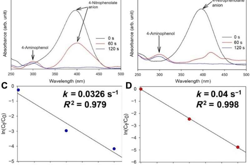
In this way, Iaroslav Petrenko and co-workers developed catalytically active, biomimetic materials using natural feedstock. They engineered centimeter-scale, mechanically stable carbon materials with controlled 3-D microarchitecture, using collagen matrices in a hybrid carbonization process and coated the spongin thermolysis products with copper. The researchers maintained the fine surface of 3-D carbon after functionalization with Cu/Cu2O for the resulting CuCSBC product. The product showed exceptional potential and stability in simulated sea water at 50C and in deionized water. The team formed a renewable and stable biomimetic CuCSBC catalyst to remove 4-NP from contaminated marine environments. The materials engineering technique is economically feasible; to farm and cultivate spongin and form mechanically robust, carbonized versions in the lab. Future research will focus at the atomic scale of the materials architecture to provide further insight to form optimized and more efficient bioinspired materials.
More information: Iaroslav Petrenko et al. Extreme biomimetics: Preservation of molecular detail in centimeter-scale samples of biological meshes laid down by sponges, Science Advances (2019). DOI: 10.1126/sciadv.aax2805
Shengjie Ling et al. Nanofibrils in nature and materials engineering, Nature Reviews Materials (2018). DOI: 10.1038/natrevmats.2018.16
Leszek Nikiel et al. Raman spectroscopic characterization of graphites: A re-evaluation of spectra/ structure correlation, Carbon (2003). DOI: 10.1016/0008-6223(93)90091-N
Journal information: Science Advances , Carbon
© 2019 Science X Network


