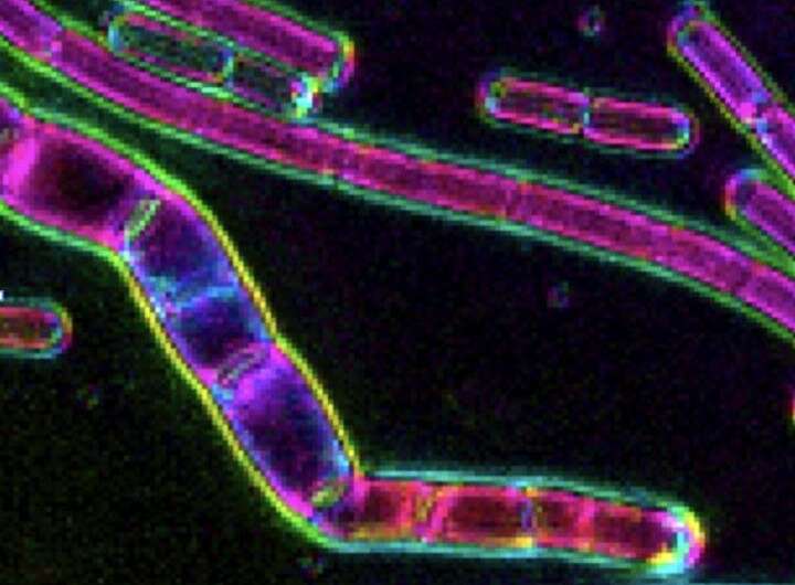
Staying in shape: MBL microscopy helps reveal how bacteria grow long, not wide

The slender, rod-shaped Bacillus subtilis is one of the best-studied bacteria in the world, a go-to system for exploring and understanding how bacteria grow, replicate and divide. One of its outstanding mysteries has been how it manages to keep its precise diameter while growing and and getting bigger end-to-end.
This week, a team led by Ethan Garner of Harvard University describes the opposing and balanced enzymatic actions that keep B. subtilis from bulging wide while it builds up its inner cell wall and elongates. The study, in Nature Microbiology, is a collaboration with microscopy developer Rudolf Oldenbourg of the Marine Biological Laboratory (MBL).
"I had been impressed by Rudolf's work for many years and always hoped that I (or someone) would introduce polarization microscopy to bacterial cell biology," Garner says. This paper was his opportunity.
With polarization microscopy, scientists can visualize the orientation of individual molecules in a live cell, and how that orientation may change over time. "Polarization microscopy was key to this project," Garner says, giving his team essential and hard-to-obtain information on the orientation of material that B. subtilis adds to its cell wall as it grows.
"As I have been giving talks on this work, the bacterial community has been incredibly impressed by this [polarization microscopy] assay," Garner says. "There are many other bacteria that people want to explore with it."
Oldenbourg, a senior scientist at MBL, is happy to oblige. "We are standing ready to support the bacteria research community through the OpenPolScope Resource at MBL," he says.
More information: Michael F. Dion et al, Bacillus subtilis cell diameter is determined by the opposing actions of two distinct cell wall synthetic systems, Nature Microbiology (2019). DOI: 10.1038/s41564-019-0439-0
Journal information: Nature Microbiology
Provided by Marine Biological Laboratory


