Molecular Velcro helps illuminate DNA repair

Using a piece of molecular "Velcro" to attach a light-emitting probe to a protein molecule, University of Wollongong (UOW) researchers have unlocked the mystery of how an important protein goes about repairing damaged DNA in bacteria, with implications for understanding how antibiotic resistance develops.
Much like how an emergency services hub coordinates various emergency response teams, the protein RecA leaps into action when DNA is damaged. RecA sounds an SOS alarm, with more than 40 different genes responding to the call to action. It assesses the extent of the damage and what repairs are needed and coordinates the repair activity at the damage site.
However, the repair process can generate mutations, said Molecular Horizons Research Fellow and study lead author Dr. Harshad Ghodke. While RecA activates or switches on the various repair mechanisms that don't introduce errors, it also switches on mechanisms that introduce errors while replicating DNA as a last-ditch attempt to help cells survive. Unfortunately, this process can lead to critical changes in the DNA sequence.
"These changes or mutations are no longer recognised as errors, and the new sequence is replicated in new generations of cells. It doesn't revert back to its original form," Dr. Ghodke said.
This creates a problem for treating bacterial infections. While antibiotics go to work to kill the bacteria, RecA swoops in to help cells survive the antibiotic treatment, and the cells that survive now have potentially antibiotic resistant mutations that render drugs ineffective.
The difficulty for researchers thus far in understanding how RecA does its job, and potentially designing drugs that counter its repair work, is that no one has been able to see where exactly repair activities occur inside living cells.
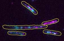
"RecA surrounds a single strand of DNA to form a filament that then signals the SOS response," Dr. Ghodke said. "Typically, researchers would attach a bright fluorescent tag to RecA so they can take images of it as it goes to work. But with the attached tag, the RecA doesn't do its job very well, and stops functioning as it would in the cellular environment."
The fluorescence signal from the tag also makes it difficult to distinguish RecA that is actively involved in repair work from that which is idle or stored away in the cell awaiting an emergency call out.
"Imagine taking an aerial photo of a city where you can only see fire trucks directly below. You can't tell if they are actively fighting fire or waiting for a call in the fire station. If we only wanted to see the fire trucks at the site of a burning building, you could attach lights to fire hydrants so that they turn on when fire trucks attach to them, and conclude that adjacent buildings are on fire.
"We did a similar thing with visualising the RecA filament. We used a viral protein that naturally interacts with the RecA filament so it wouldn't interfere with how it works, while lighting up the RecA filament as it takes part in the damage response."
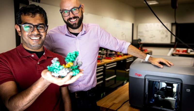
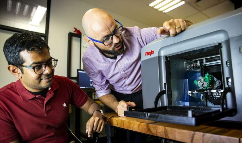
To help visualise this radical new approach to illuminate this DNA repair process inside living Escherichia coli bacteria cells, the researchers turned to 3-D printing to create physical models of RecA to enable them to see its shape and form.
Molecular Horizons Director, Distinguished Professor Antoine van Oijen, said a key to biological processes is to think in structures and shapes.
"We know from the imaging we do that proteins are dynamic objects. If we think of them as 3-D structures we can start to visualise how they change and what causes those changes, leading to a clearer understanding of how these proteins work.

"With a physical structure, you can see the interfaces and design methods to attach other proteins. Then using sophisticated imaging tools we can take short films that for the first time, really show us how they work."
Professor van Oijen said the critical piece of the puzzle the team has solved paves the way for new drug treatments that overcome antibiotic resistance.
In some cases, mutated cells deactivate the drug or no longer have the target protein the drug molecules are searching for and a rogue cell is not destroyed.

"Antibiotic resistance is a hugely important global challenge. We don't want to get rid of antibiotics altogether because when they work they're incredibly effective. If we can visualise these processes we can then understand the physical connections between molecules and the structure of proteins and potentially design new drugs that will prevent bacterial cells from becoming resistant."
The research was published in the journal eLife.
More information: Harshad Ghodke et al. Spatial and temporal organization of RecA in the Escherichia coli DNA-damage response, eLife (2019). DOI: 10.7554/eLife.42761
Journal information: eLife
Provided by University of Wollongong


