A novel approach for the study of integral membrane proteins

The membranes that surround our cells contain a large number of proteins. Membrane proteins are therefore a crucial class of macromolecules in living systems. They play key roles, such as providing transport gateways into and out of the cell, facilitating signalling between cells, as well as being involved in enzyme catalysis. These functional roles make them particularly important as drug targets, with the majority of current therapeutics targeting membrane proteins.
However, structural studies of integral membrane proteins (IMPs) have proved extremely challenging, since most of them are difficult to study properly in the absence of their lipid environment. This often prevents them from being crystallised – a method commonly used in classical structural biology. Alternative approaches are therefore required for structural studies of IMPs in membranous environments. For this purpose, the Life Sciences Group at the Institut Laue-Langevin (ILL), in collaboration with Copenhagen University, has successfully pioneered the development of stealth carrier nanodiscs. In this approach, a sophisticated deuterium labelling method is used to make the membrane effectively invisible to low-resolution neutron diffraction while still highlighting the structure of IMPs within their usual lipidic environment, as published in Acta Crystallographica D in 2014.
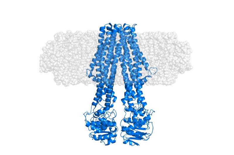
More recently, the first structural study of an integral membrane protein using this stealth carrier nanodisc deuteration strategy has just been completed. This was carried out using the Deuteration Laboratory (D-Lab) platform of the Partnership for Structural Biology (PSB) in conjunction with small-angle neutron scattering (SANS) and X-ray scattering (SAXS) provided through the PSB SANS/SAXS platform. As published in the journal Structure by Josts et al, the international team, led by Henning Tidow, University of Hamburg, applied this method to an ATP-binding cassette (ABC) transporter protein, MsbA – which plays an important role in lipid transport in bacteria. The resulting neutron scattering data, mostly acquired using the D11 instrument at ILL, allowed direct observation of the signal from the solubilised membrane protein without contribution from the surrounding lipid. The SAXS data provided a clear reference for the external shape of the nanodisc, inclusive of the lipid bilayer.
In addition, conformational changes in MsbA were studied, demonstrating the sensitivity of the method and its general applicability to structural studies of IMPs.
This approach is likely to become increasingly important in future studies of these difficult, but critically important, biological macromolecules, in turn supporting a better understanding for the development of drugs aimed at membrane proteins.
More information: Inokentijs Josts et al. Conformational States of ABC Transporter MsbA in a Lipid Environment Investigated by Small-Angle Scattering Using Stealth Carrier Nanodiscs, Structure (2018). DOI: 10.1016/j.str.2018.05.007
Journal information: Structure
Provided by Institut Laue-Langevin


