Researchers aid groundbreaking test of serial crystallography technique

Rice University scientists used a rapidly pulsing X-ray laser to show how drug-resistant tuberculosis bacteria deactivate the antibiotic molecules intended to treat the deadly lung disease.
Rice biochemist George Phillips and graduate student and co-lead author Jose Olmos are part of the National Science Foundation-supported BioXFEL Center that captured the groundbreaking proof-of-principal results through a technique called mix-and-inject serial crystallography.
To do so required the use of a new tool, an X-ray free-electron laser (XFEL) that promises a serious upgrade to the painstaking, century-old process of characterizing molecules through X-ray spectroscopy. The laser is located at the Department of Energy's SLAC National Accelerator Laboratory at Stanford University.
Defining molecular structures is key to understanding how they function, Phillips said. The new discovery demonstrates scientists' rapidly developing ability to take snapshots of dynamic biological processes as they happen.
True to its name, the mix-and-inject technique feeds a narrow stream of crystalized molecules in a solution to the laser. When the laser hits a crystal with a 20-femtosecond (quadrillionth of a second) pulse, it obliterates the crystal – but not before producing a diffraction pattern on a detector that shows the molecule's atomic structure.
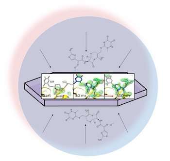
In an open-access paper in BMC Biology, the researchers led by Marius Schmidt, a professor at the University of Wisconsin-Milwaukee,described mixing the antibiotic ceftriaxone with a resistant enzyme used by bacteria, beta-lactamase, and feeding it to the pulsing laser. Because they could adjust the time between mixing and arrival at the laser, they captured diffraction patterns of the crystallized molecules not only in random orientations but also at several stages of interaction.
"While there have been elegant studies to observe protein motions with light-induced changes, our work illustrates that a larger class of proteins, namely enzymes, can be investigated in a time-resolved fashion at LCLS (Linac Coherent Light Source)and other XFELs," Olmos said.
Phillips said the experiment proved XFEL's utility to capture diffraction patterns from crystals a millionth of a meter across or less, much smaller than previous techniques. "This will teach us more about how nature has selected for and designed these molecules to work," he said. "It's not unlike seeing a bicycle being pedaled: You get more than a static picture and a better understanding of how it works.
"Anytime you want to engineer a protein or recreate a molecular machine, knowing more about how they work at a fundamental level is going to be helpful, whether it's breaking down cellulose for biofuels or designing a new drug or improving an existing drug."
In his presentations, Phillips compares the ability to take snapshots of proteins in action to 19th- century images by Eadweard Muybridge that captured the midstride motion of a galloping horse. (By coincidence, the horse was owned by Stanford's founder.)
The researchers expect the soon-to-be upgraded XFEL at Stanford, a new facility in Europe and others in the works around the world will allow scientists to capture structures in minutes rather than days and give them more detailed data about chemical processes.
Phillips has high hopes the upgraded tools will also help capture the structures of molecules without having to crystallize them first.
"If we can get the X-ray beam and the background scattering small enough and the readout beam clean enough, then in theory, instead of parading crystals, we could parade single molecules into the laser to build up diffraction patterns," he said.
"The Stanford laser right now fires at 100 hertz (cycles per second)," Phillips said. "The European XFEL is going to fire at 10,000 hertz. That's quite an upgrade because it gives us a lot more chances to hit the molecules as they stream in."
He said the center ultimately hopes to capture structural data for molecular reactions on the fly.
"It could be two proteins coming together and learning to recognize each other, it could be the interaction of a virus with an antibody, it could be the interaction of some substrate with an enzyme or anything you can do by mixing or with external stimulation," Phillips said. "Once you can do that, the sky's the limit."
More information: Christopher Kupitz et al. Structural enzymology using X-ray free electron lasers, Structural Dynamics (2016). DOI: 10.1063/1.4972069
Allen M. Orville. Entering an era of dynamic structural biology…, BMC Biology (2018). DOI: 10.1186/s12915-018-0533-4
Journal information: BMC Biology
Provided by Rice University


