Multiple optical measurements reveal the single cell activation without contrast agent

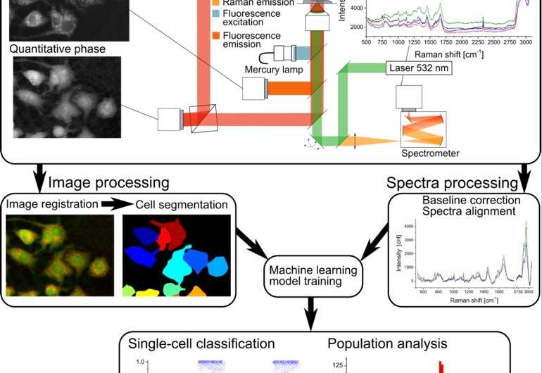
Nicolas Pavillon (Assistant Professor), Nicholas I. Smith (Associate Professor, Immunology Frontier Research Center, Osaka University) and collaborators developed a label-free multimodal microscopy platform that allows the non-invasive study of cellular preparations without the need of any additional chemicals or contrast agent. The parameters extracted from these measurements, coupled with machine algorithms, enable the study of fine cellular processes such as macrophage cells activation upon exposure to lipopolysaccharide (LPS). The authors demonstrate that activation, as well as partial activation inhibition, can be observed at single-cell level through phenotypic and molecular characterization purely through non-invasive optical means.
Label-free optical measurements have become popular thanks to their ability to observe samples non-invasively. Techniques such as quantitative phase microscopy or Raman spectroscopy, as used in this study, have been extensively used in recent years to characterize specimens and identify cells from different origins. However, the specificity of these approaches usually only allows for the discrimination of cell types. The group shows in this study that fine processes such as macrophage activation within populations of identical cells is also possible.
Standard analysis methods are often destructive to the sample, or rely on contrast agents to detect specific molecules of interest, which limits the study to known target molecules. The approach developed here is based on non-invasive techniques that rely on the endogenous contrast of the sample, i.e. on its phenotype and whole intracellular molecular content. Furthermore, standard assays, despite their sensitiveness, often measure samples at a population level, taking advantage of the cumulative effect on secreted molecules, but losing the information about individual cellular response. The presented approach provides measurements at single-cell level, enabling the study of cell-to-cell variability within populations.
More information: Nicolas Pavillon et al. Noninvasive detection of macrophage activation with single-cell resolution through machine learning, Proceedings of the National Academy of Sciences (2018). DOI: 10.1073/pnas.1711872115
Journal information: Proceedings of the National Academy of Sciences
Provided by Osaka University


