Intracellular transport in 3-D

Ludwig Maximilian University researchers have visualized the complex interplay between protein synthesis, transport and modification.
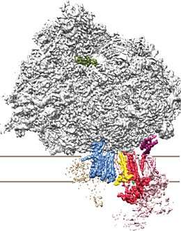
Cells of higher organisms are densely populated by a tubular network of membranes known as the endoplasmic reticulum (ER). ER-resident complexes serve as a binding platform for ribosomes, the cellular machines responsible for protein synthesis. ER-attached ribosomes synthesize proteins destined for various intra- and extracellular locations. To warrant successful delivery, many of these proteins are chemically modified with the molecular equivalent of an address tag upon passage through the ER membrane. LMU researchers led by Professor Roland Beckmann at the Gene Center, in collaboration with colleagues at the Max Planck Institute for Biochemistry, have now determined the three-dimensional structure of the macromolecular complex which couples this labeling-step to ribosomal protein synthesis and protein transport at the ER membrane. Their study provides the basis for a detailed understanding of this vital cellular process and has been published in the journal Science.
All biological membranes are made up of a phospholipid bilayer, which is essentially impermeable to polar (electrically charged) molecules such as proteins. However, certain specialized proteins can act as channels for the passage of other proteins through lipid bilayers. At the ER membrane such a 'translocon' allows proteins synthesized by ER-bound ribosomes to enter the interior of the ER or to be integrated into the ER membrane, respectively. As it passes across the membrane, the growing protein is modified at specific sites by the attachment of an 'oligosaccharide' made up of a chain of 14 sugar molecules, in a process known as glycosylation. This chemical label ensures that the protein is subsequently delivered to the correct destination. In addition, the label plays a vital role in enabling the nascent protein to fold into the appropriate shape required for its biological function. "Errors in glycosylation lead to the accumulation of improperly folded proteins, and these in turn activate cellular stress responses – often with fatal consequences for the cell," says Katharina Braunger, a member of Beckmann's team and first author of the new study.
The enzyme complex which attaches the oligosaccharide as the nascent protein chain is fed through the translocon by the ribosome is known as the OST (oligosaccharyltransferase) complex. In higher organisms the OST complex is present in two different compositions. Using a combination of cryo-electron microscopy and -tomography, Beckmann and colleagues have now obtained structural evidence that these two forms also differ in their function. A-form OST interacts with both the active ribosome and the protein conducting channel to form a stable complex, and it modifies the growing protein during its production. In contrast, the other variant is unable to bind to the translocon. "The B variant is responsible for proof reading as well as the modification of sites that are not accessible to the A variant," says Beckmann. The new data has allowed the team to define the three-dimensional organization of the subunits in ribosome-bound OST complexes, and to develop a molecular model for their function. The model allows them to propose the basis for differences in specificity between the two variants, and explains how protein transport and glycosylation are coupled mechanistically.
More information: Katharina Braunger et al. Structural basis for coupling protein transport and N-glycosylation at the mammalian endoplasmic reticulum, Science (2018). DOI: 10.1126/science.aar7899
Journal information: Science
Provided by Ludwig Maximilian University of Munich


