A microscope within a microscope

No single microscope can image all aspects of a sample at the same time and so the use of two or more imaging methods to study a sample - correlative imaging - is common-place.
Lucy Collinson leads the Electron Microscopy Science Technology Platform at the Francis Crick Institute. The platform provides expertise in imaging the structures of molecules, cells and tissues at high resolution.
Correlative imaging has enabled Crick researchers working in collaboration with Lucy's team to study how synapses in the brain form, the early processes in production of blood-clotting proteins and how HIV particles are released from infected cells.
Now, by collaborating with instrument manufacturers, Lucy's team have taken the technology a step further.
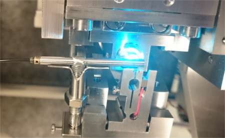
They have developed a new way to image structures that overcomes the limitations of existing technology. They have done this by putting a fluorescent light microscope within an electron microscope.
"One major challenge in correlative imaging is to prepare the sample in a way that it can be imaged in both the light and electron microscope," explains Lucy. "We developed a protocol that preserves the fluorescent proteins that light up under the light microscope, while staining and embedding the sample so that cells can be seen in the electron microscope and withstand the vacuum environment. This allows us to image the samples in one machine - an integrated light and electron microscope."

Imaging a sample in this way reveals the dynamic function of proteins (revealed by the light microscope) within the context of the cell and tissue structure (shown in great detail by the electron microscope).
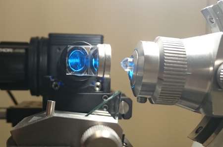
Martin Jones and Lizzy Brama, two physicists within the Microscopy Prototyping group, designed and built a 'miniLM,' similar in concept to fluorescence endoscopy systems used in fluorescence-guided surgery.
They also designed a second system, the 'ultraLM,' which aids in sample preparation of the fluorescent resin-embedded cells in the ultramicrotome tool that slices samples into sections.
Development work took place at the Crick, with the idea that it should be made available to the community as open-source technology. The aim is for any facility that has the skills to be able to build and retrofit these miniaturised fluorescence microscopes into their own electron microscopes. There are also plans to licence the technology to commercial manufacturers.
Provided by The Francis Crick Institute


