3D printed frog skeletons for classrooms

Scientists from Massey University have developed a simple 3D scanning and printing method that will help students learn anatomy.
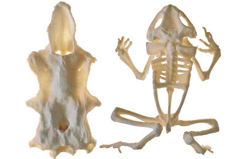
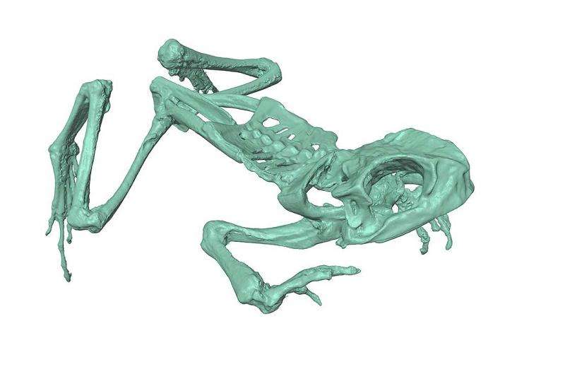
3D digital replicas of a cane toad skeleton and the tough cartilage from the head of a spiny dogfish were made using consumer-level scanners. The skeleton and cartilage replicas were printed using a selective laser sintering 3D printer. These test cases explain how high-quality replicas can be made more accessible and make a case for wider application of 3D printing in anatomy.
Lead author Dr Daniel Thomas of Massey's Institute of Natural and Mathematical Sciences says the aim was to make anatomy more accessible to students and teachers.
"Anatomy teaches us about the ecology and evolution of an animal and can give us crucial information for developing conservation strategies. It's not always possible for learners to study original anatomy specimens though, which is where high-quality 3D printed models come in.
"Imagine a classroom in Silverdale being able to print a moa skeleton or a university class in America being able to examine a kakapo beak that was scanned here in New Zealand."
The School of Engineering and Advanced Technology printed the pieces using their laser sintering 3D printer. "The scanning system we used is reasonably inexpensive for a school or university to buy. Online services for 3D printing are great if an educator or learner doesn't have their own printer.

"There is no maximum size limit for printing or scanning, as bones that are larger than the printing chamber can be printed in multiple pieces," says Dr Thomas.
He acknowledges anatomical models don't account for biological variation and there are aspects lost by moving away from dissection. However, the fundamental advantage of models is they can provide educational opportunities to learners who may otherwise not have access to original specimens.
Dr Thomas will use 3D scanning and printing to increase the range of specimens that students can study in his vertebrate zoology classes.
The frog skeleton and dogfish cartilage models can be used to make large class sets and are available for downloading and 3D printing from the NZ Fauna website. Click here for frog skeleton and click here for the spiny dogfish.
Provided by Massey University


