Ancient mummies meet modern technology

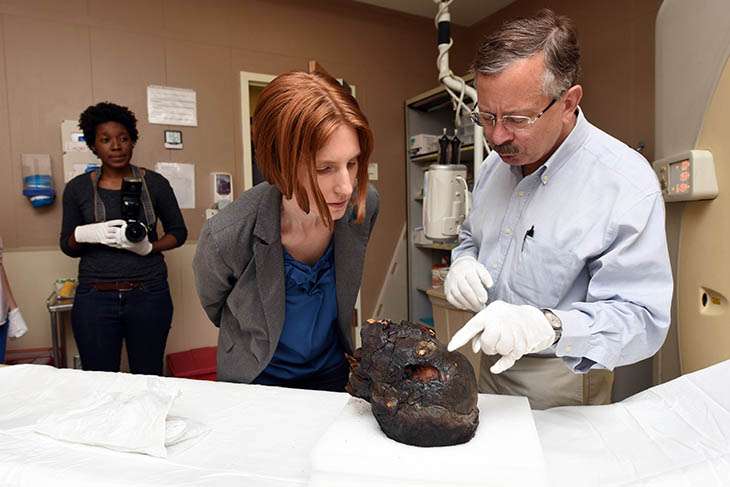
For her senior honors thesis, Tulane University student Savanna Bailey accompanied two ancient Egyptian mummies—well, actually, just their heads—to Tulane Medical Center in downtown New Orleans. The patients in question hadn't had a medical appointment in 18 years, so the trip was quite a milestone.
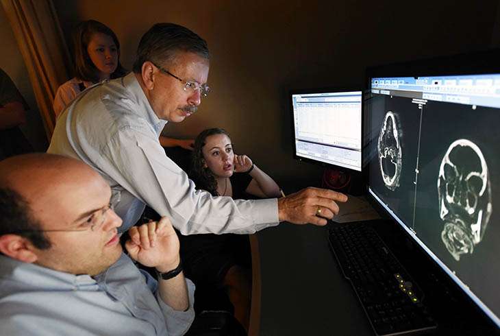
Bailey, along with Tulane anthropologists and graduate students, stared open-mouthed at the brain scans produced by the hospital's high-tech CT (computerized tomography) equipment.
"These long-dead individuals reveal much about how they lived, little to nothing about how they died, and much about their mummification and preservation since they were alive," Bailey wrote in her thesis, which won the approval of her faculty committee.
Bailey is a student of anthropology professor John Verano. Near Verano's office in Dinwiddie Hall is the resting place of the two mummies that were donated to Tulane in 1852.
Although the mummies were scanned in 1998, today's newer equipment "has come a long way," Verano said," and that's one of reasons we decided to do this, to get better material." Dr. Scott Beech, head of the TMC radiology department, worked with Verano to arrange the scans.
Only the heads, which were already unattached, were scanned to avoid moving the fragile, 3,000-year-old mummies, Verano said.
Analysis of the female mummy's teeth showed her to be between 13.5 and 15.5 years old at death, verifying previous research. Scans of the male mummy, a priest and overseer of craftsmen at one of the most important temples in Egypt, revealed that he was more than 50 years old, based on an extensive amount of tooth wear.

Anthropology research associate and Egyptologist Melinda Nelson-Hurst said that despite the male mummy's high status, "he was likely considerably uncomfortable by the time of his death, due to dental abscesses and degeneration of the cervical vertebrae (in his neck)."
While tomb and temple artwork typically show ancient Egyptians as young and healthy, Nelson-Hurst said studies such as these "broaden our picture of life (and death!) in ancient Egypt." The use of technology such as CT scanning provides "a vital balance" to other information from archeological remains and ancient art and texts.
Provided by Tulane University


