A novel liquid-immersible micro-electromechanical systems scanning mirror

Dr. Jun Zou, an associate professor in the Department of Electrical and Computer Engineering at Texas A&M University, and several co-authors recently had their paper published in the prestigious research publication Nature Methods.
Zou and a Ph.D. student under his supervision, Chih-Hsien Huang, were among the authors of the research paper titled "High-speed label-free functional photoacoustic microscopy of mouse brain in action." It is based on a four-year joint work between Zou's Micro Imaging/Sensing Devices and Systems Lab in the department and Dr. Lihong Wang's Optical Imaging Lab at the Washington University in St. Louis. Their work was funded by the National Science of Foundation and the National Institutes of Health.
Zou's team first conceived and developed a novel liquid-immersible MEMS (micro-electromechanical systems) scanning mirror. The unique feature of this MEMS scanning mirror is its capability of fast and simultaneous scanning of both focused optical and high-frequency ultrasound beams in a liquid environment, which is necessary for high-frequency ultrasound propagation. It overcomes the major limitations in existing MEMS scanning mirrors, which can only operate in air, but will fail in liquids.
With the new water-immersible MEMS scanning mirror, the two teams worked together to build a new scanning microscope to enable high-speed and high-resolution photoacoustic imaging on the intact brain of live mice. Photoacoustic imaging is a hybrid biomedical imaging modality, which utilizes optical and ultrasound waves to interrogate biological cells and tissues even without additional contrast agents.
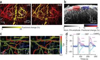
The new scanning photoacoustic microscope achieved 50-times higher speed and 5-times higher resolution than its predecessors. This makes it possible to capture not only still snapshots of the vascular network of the mouse brain, but more importantly blood flow dynamics and oxygen metabolism at capillary level. In this work, the vascular morphology, blood oxygenation, blood flow and oxygen metabolism in both resting and stimulated states in the mouse brain were revealed. This work provides a new approach to study the complex neurological functions of the brain. Given the importance of oxygen metabolism in basic biology and diseases progression, it is also expected to find broad applications from fundamental biomedical researches to clinical diagnosis.
More information: "High-speed label-free functional photoacoustic microscopy of mouse brain in action" Nature Methods (2015) DOI: 10.1038/nmeth.3336
Journal information: Nature Methods
Provided by Texas A&M University


