Improving cancer treatment through better imaging technologies
Since January 2014, the HELICOID project has been developing hyperspectral imaging technologies capable of differentiating between healthy and diseased tissues. The February 2015 meeting in Brussels enabled project partners to meet and discuss the progress that has been made.
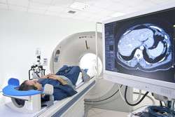
Hyperspectral imaging – also known as imaging spectroscopy – works by generating very high-dimensional images through the use of sensor optics. This imaging technique provides researchers and medical staff with much more detailed information than traditional imaging techniques can, thus aiding in the removal of tumours.
One area that HELICOID researchers are especially interested in is brain tumours, which, more than any other cancers, can resemble the normal surrounding tissue. This makes identifying and operating on brain tumours extremely difficult.
As a result, although malignant primary brain tumours are the 13th most frequent cancers in adults, they are the fifth most common cause of cancer death for those under 65, due to poor prognosis. Moreover they are the second most common cancer and the most common cause of cancer death in children.
The Brussels meeting shared information about the project's achievements to date. In November, the first prototype of the HELICOID hyperspectral image system was completed on time. This system has now been deployed to a consortium of hospitals and should begin delivering hyperspectral datasets to the team in the next few months.
The project website has also been launched. This fulfils two key roles: providing relevant and accessible information about the project to the general public and the research community, and providing project partners with a convenient means of exchanging key information.
These developments have enabled the HELICOID consortium to make a start on the challenging task of brain cancer detection. Using the hyperspectral signatures of healthy tissues and cancerous tissues, a model of how cancer affects the hyperspectral signature is being developed. This technique will aid surgeons in identifying exactly what tissues must be removed.
Other types of tumours will shortly be analysed, including lung and breast cancers. These represent the two most common cancers in the world. As cancer results in a change in cellular physiology, the HELICOID project will detect this as a change in the hyperspectral signature. The team will try to determine whether there is a certain pattern that can be identified as a cancer hyperspectral signature. In addition, high-efficiency hardware and software will be developed with the aim of recognising cancer tissues in real time.
The HELICOID project represents a potential watershed in cancer detection. At present, the main tool for differentiating between normal and malignant tissue remains the human eye. Other techniques have been developed but none has succeeded in reliably differentiating tissue.
By providing a precise definition of the edges of malignant tissue in real time, hyperspectral imaging promises to speed up diagnosis and surgery and save lives. The HELICOID project is due for completion in December 2016.
More information: For further information please visit HELICOID: www.helicoid.eu/


