November 27, 2014 report
Researchers find a way to improve FELs approach used to study Photosystem II in plants

A team of researchers in Japan has developed a technique for improving the free electron lasers (FELs) approach to studying Photosystem II that allows for creating high-resolution images of the oxygen evolving complex (OEC) in plants. In their paper published in the journal Nature, the team describes their new technique and how their work might lead to a better understanding of the entire photosynthesis process. Ilme Schlichting, of the Max Planck Institute offers a News & Views piece on the work done by the team in the same journal issue.
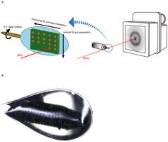
Scientists have been studying plants for quite some time trying to understand how photosynthesis works, and while they have made a great deal of progress, there are still some parts that aren't completely understood, one of those is Photosystem II, the initial reaction in photosynthesis, where a plant uses energy from light to split water molecules into oxygen and hydrogen atoms. The problem lies in catching the plant mechanism in action and taking a picture of it. Scientists have used lasers in the past, but that causes damage to the plant parts being studied. More recently, X-ray free electron lasers (FELs) have been developed that allow for a technique called serial femtosecond crystallography—where a sample is exposed to a small X-ray pulse, over and over, resulting in the creation of an image that can be used for study. But, it still doesn't allow for the high resolution (because of diffraction issues) scientists really want. In this new effort, the researchers have taken the FELs approach a step further and in so doing have come up with a way to create the high resolution images scientists have been hoping for.
The team brought with them expertise in dehydration techniques that allow for slow, controlled large crystal growth—adding such a process to the FELs approach allowed for creating very high resolution images of Photosystem II (in its dark resting state) for the first time. Schlichting notes that because the team used large crystals, the technique also allows for using more than one diffraction pattern by moving the sample around, thus giving researchers more views.
In addition to helping to learn more about how plants use the sun's energy to perform photosynthesis, scientists may be able to use the new technique to develop better man-made versions of the OEC to hopefully offer a new type of solar cell that will finally get us off burning fossil fuels.
More information: Native structure of photosystem II at 1.95 Å resolution viewed by femtosecond X-ray pulses, Nature (2014) DOI: 10.1038/nature13991
Abstract
Photosynthesis converts light energy into biologically useful chemical energy vital to life on Earth. The initial reaction of photosynthesis takes place in photosystem II (PSII), a 700-kilodalton homodimeric membrane protein complex that catalyses photo-oxidation of water into dioxygen through an S-state cycle of the oxygen evolving complex (OEC). The structure of PSII has been solved by X-ray diffraction (XRD) at 1.9 ångström resolution, which revealed that the OEC is a Mn4CaO5-cluster coordinated by a well defined protein environment1. However, extended X-ray absorption fine structure (EXAFS) studies showed that the manganese cations in the OEC are easily reduced by X-ray irradiation2, and slight differences were found in the Mn–Mn distances determined by XRD1, EXAFS and theoretical studies. Here we report a 'radiation-damage-free' structure of PSII from Thermosynechococcus vulcanus in the S1 state at a resolution of 1.95 ångströms using femtosecond X-ray pulses of the SPring-8 ångström compact free-electron laser (SACLA) and hundreds of large, highly isomorphous PSII crystals. Compared with the structure from XRD, the OEC in the X-ray free electron laser structure has Mn–Mn distances that are shorter by 0.1–0.2 ångströms. The valences of each manganese atom were tentatively assigned as Mn1D(III), Mn2C(IV), Mn3B(IV) and Mn4A(III), based on the average Mn–ligand distances and analysis of the Jahn–Teller axis on Mn(III). One of the oxo-bridged oxygens, O5, has significantly longer distances to Mn than do the other oxo-oxygen atoms, suggesting that O5 is a hydroxide ion instead of a normal oxygen dianion and therefore may serve as one of the substrate oxygen atoms. These findings provide a structural basis for the mechanism of oxygen evolution, and we expect that this structure will provide a blueprint for the design of artificial catalysts for water oxidation.
Journal information: Nature
© 2014 Phys.org


