Handheld scanner could make brain tumor removal more complete, reducing recurrence

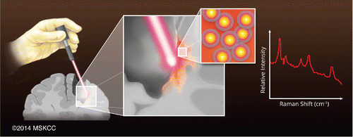
Cancerous brain tumors are notorious for growing back despite surgical attempts to remove them—and for leading to a dire prognosis for patients. But scientists are developing a new way to try to root out malignant cells during surgery so fewer or none get left behind to form new tumors. The method, reported in the journal ACS Nano, could someday vastly improve the outlook for patients.
Moritz F. Kircher and colleagues at Memorial Sloan Kettering Cancer Center point out that malignant brain tumors, particularly the kind known as glioblastoma multiforme (GBM), are among the toughest to beat. Although relatively rare, GBM is highly aggressive, and its cells multiply rapidly. Surgical removal is one of the main weapons doctors have to treat brain tumors. The problem is that currently, there's no way to know if they have taken out all of the cancerous cells. And removing extra material "just in case" isn't a good option in the brain, which controls so many critical processes. The techniques surgeons have at their disposal today are not accurate enough to identify all the cells that need to be excised. So Kircher's team decided to develop a new method to fill that gap.
The researchers used a handheld device resembling a laser pointer that can detect "Raman nanoprobes" with very high accuracy. These nanoprobes are injected the day prior to the operation and go specifically to tumor cells, and not to normal brain cells. Using a handheld Raman scanner in a mouse model that mimics human GBM, the researchers successfully identified and removed all malignant cells in the rodents' brains. Also, because the technique involves steps that have already made it to human testing for other purposes, the researchers conclude that it has the potential to move readily into clinical trials. Surgeons might be able to use the device in the future to treat other types of brain cancer, they say.
More information:
"Guiding Brain Tumor Resection Using Surface-Enhanced Raman Scattering Nanoparticles and a Hand-Held Raman Scanner" ACS Nano, Article ASAP
DOI: 10.1021/nn503948
Abstract
The current difficulty in visualizing the true extent of malignant brain tumors during surgical resection represents one of the major reasons for the poor prognosis of brain tumor patients. Here, we evaluated the ability of a hand-held Raman scanner, guided by surface-enhanced Raman scattering (SERS) nanoparticles, to identify the microscopic tumor extent in a genetically engineered RCAS/tv-a glioblastoma mouse model. In a simulated intraoperative scenario, we tested both a static Raman imaging device and a mobile, hand-held Raman scanner. We show that SERS image-guided resection is more accurate than resection using white light visualization alone. Both methods complemented each other, and correlation with histology showed that SERS nanoparticles accurately outlined the extent of the tumors. Importantly, the hand-held Raman probe not only allowed near real-time scanning, but also detected additional microscopic foci of cancer in the resection bed that were not seen on static SERS images and would otherwise have been missed. This technology has a strong potential for clinical translation because it uses inert gold–silica SERS nanoparticles and a hand-held Raman scanner that can guide brain tumor resection in the operating room.
Journal information: ACS Nano
Provided by American Chemical Society


