Evidence that disproves a long-standing assumption about fish development gives insight into evolution of skeletons

A Singapore-based research team has used fluorescent labeling of embryonic cell populations to pinpoint the origin of scales and fins in modern-day fish. These tissues are evolutionary relics of the first skeletons and were widely assumed to originate from an embryonic cell population known as the 'trunk neural crest'. Now, research led by Tom Carney of the A*STAR Institute of Molecular and Cell Biology has shown that scales and fins actually develop from a cell population called the mesoderm.
In both mammals and fish, two embryonic cell populations contribute to the formation of the skeleton—the cranial neural crest generates most of the skull and jaw bones, and the mesoderm generates the remainder of the skeleton. In mammals, trunk neural crest cells, a third population, do not contribute to skeletal development, but previous cell-labeling studies suggested that they did in ancient vertebrates and still do in some modern animals. Despite inaccurate cell labeling in these studies, the widely held assumption remained. However, no-one had directly demonstrated the role of these trunk neural crest cells.
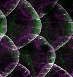
Carney and his team labeled embryonic cell populations with fluorescent molecules to determine which populations contribute to the fin skeleton and scales in modern fish. New cells originating from these populations also expressed the label, allowing the team to see which cells in the body developed from each population.
"We found that fish scales and fin rays derive entirely from the mesoderm, with no [trunk] neural crest contribution," explains Carney. "This [finding] indicates that the mesoderm can generate this ancient type of bone and suggests that when skeletons first evolved, they developed from the mesoderm."
The team's discovery is contrary to the previous assumption about the role of the trunk neural crest in skeleton development. They therefore investigated further using genetic manipulation and more cell labeling.
"We went on to show that the trunk neural crest in fish does not generate any skeletal-type cells," says Carney. "This suggests that the contribution of cranial neural crest cells to the skeleton may have evolved independently in the head and has never been a general feature of the whole neural crest."
The results are likely to change our understanding of skeleton evolution. Carney says that: "Many of the important 'innovations' of the skeleton can now be considered as 'inventions' of the mesoderm, with the neural crest only playing a specific role in generating the skeleton of the head."
More information: Lee, R. T. H., Thiery, J. P. & Carney, T. J. Dermal fin rays and scales derive from mesoderm, not neural crest, Current Biology 23, R336–R337 (2013). www.cell.com/current-biology/abstract/S0960-9822%2813%2900262-5
Lee, R. T. H., Knapik, E. W., Thiery, J. P. & Carney, T. J. An exclusively mesodermal origin of fin mesenchyme demonstrates that zebrafish trunk neural crest does not generate ectomesenchyme, Development 140, 2923–2932 (2013). dev.biologists.org/content/140/14/2923
Journal information: Current Biology , Development


