Findings from quantitative analysis of chromatin structure challenge classical model of static regularity

The DNA in the human genome is organized into irregularly folded fibers during cell division, according to a recent study by a team of researchers led by Kazuhiro Maeshima of the RIKEN SPring-8 Center and RIKEN Advanced Science Institute in Japan.
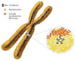
DNA is wrapped around proteins called histones to form nucleosome fibers, which are tightly compressed into the chromosomes by large protein complexes called condensins (Fig. 1). Many previous studies suggest that nucleosomes are organized into regular fiber structures that are 30 nanometers in diameter, which led to the classical model of overall chromosome structure. However, other studies suggest that these regular fiber structures exist only in specialized cell types.
To resolve these conflicting results, Maeshima and his colleagues investigated chromosome structure in mitotic, or dividing, HeLa cells using three different techniques: cryo-electron microscopy, which allows for observation of frozen, hydrated biological structures at high resolution without producing the artifacts seen in conventional EM; small-angle x-ray scattering (SAXS), which detects repeating structures in solutions of biological samples; and ultra-small x-ray scattering (USAXS), a newly developed type of SAXS that can resolve repeating structures across larger dimensions.
All three techniques produced consistent results. The researchers detected repeating structures at approximately every 11 nanometers, but not at longer distances, suggesting that chromatin is organized like beads on a string with an irregular folding pattern. The USAXS method further revealed that the chromosomes have a fractal nature: the same organization repeats itself at different scales, and that is the structures arrange into a rod shape.
Maeshima and colleagues propose two possible explanations for the rod-shaped and large-scale organization of the chromosome. One is that the condensins bind to specific sites in the DNA, causing it to form self-assembling loops that interact with each other and produce repeating structures along the axis of the chromosome. Alternatively, the formation of regularly repeating looped structures alone might be sufficient to generate the observed rod shape because of repulsive forces between adjacent loops.
The researchers predict that irregular folding would make chromosomes more flexible: this type of organization has fewer physical constraints than a regular structure. They also suggest that irregular folding is common to chromatin in interphase, or non-dividing cells, as it makes DNA more accessible to the molecular machinery for RNA transcription and DNA replication.
“We focused on chromosomes in dividing cells,” says Maeshima, “but we assume that a similar irregular folding also exists in interphase cells, and are now assessing that assumption.”
More information: Nishino, Y., et al. Human mitotic chromosomes consist predominantly of irregularly folded nucleosome fibres without a 30-nm chromatin structure. The EMBO Journal 31, 1644–1653 (2012)
Journal information: EMBO Journal
Provided by RIKEN


