Noninvasive and accurate: Absolute temperature mapping for biomedical applications
(PhysOrg.com) -- Researchers in Germany and the US have developed a new approach to magnetic resonance imaging (MRI) thermometry using encaged hyperpolarized xenon as a temperature sensor. The method allows absolute temperature mapping with an unprecedented accuracy of 0.1 C at low and ultralow sensor concentrations. The new technique, which has the potential to become a helpful tool in clinical diagnostics and therapy monitoring, is presented online in ChemPhysChem.
According to Franz Schilling, now a researcher at the Technical University of Munich and lead author of the paper, the method benefits from all the advantages of MRI including its non-invasiveness and the ability to image in any scan orientation with good spatial and temporal resolution.
Many magnetic resonance parameters are inherently temperature-sensitive; for example, proton resonance frequency (PRF), diffusion coefficient or transverse and longitudinal relaxation times. The accuracy of many conventional thermometry methods such as PRF is however limited and although such techniques provide useful relative temperature results, MRI thermometry based on encapsulated hyperpolarized xenon appears to be a more promising technology for in vivo applications because it can be used to map absolute temperatures.
Hydrogen is the most frequently imaged nucleus in MRI, but any nucleus with a net nuclear spin can potentially be imaged, including xenon (129Xe). Such gaseous isotopes must be hyperpolarized before use as their net magnetization is too low to yield a good signal under normal conditions. Schilling and co-workers have introduced xenon sensors as a temperature contrast agent for MRI thermometry to gain both high accuracy and sensitivity. They have achieved this by hyperpolarization through spin exchange optical pumping, which increases the equilibrium net spin polarization by three to four orders of magnitude.
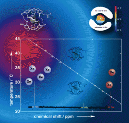
The new technique is based on the temperature-dependent chemical shift of hyperpolarized xenon in a cryptophane-A cage. This shift is linear with a slope of 0.29 ppm per °C, which is almost 30 times higher than that of the proton resonance frequency that is currently used for MRI thermometry. According to Schilling, this new direct mapping concept allows absolute temperature mapping with a previously unmatched accuracy of 0.1˚C at a sensor concentration of 150 µM. But the researchers have also demonstrated an indirect temperature detection technique, via chemical exchange saturation transfer of hyperpolarized xenon (Hyper-CEST, introduced previously by co-author Leif Schröder who currently works at the Leibniz Institute for Molecular Pharmacology in Berlin), which makes temperature mapping with nanomolar agent concentrations as low as 10 nM possible. “This absolute temperature imaging concept offers high temperature accuracy at ultralow agent concentrations”, Schilling says. The new sensors consist of three major components: a cryptophane-A cage for hosting the xenon atom, a linker, and a peptide for sufficient water solubility.
More information: Franz Schilling, Alexander Pines, MRI Thermometry Based on Encapsulated Hyperpolarized Xenon, ChemPhysChem 2010, 11, No. 16, dx.doi.org/10.1002/cphc.201000507
Provided by Wiley


