April 22, 2014 feature
Bright lights, small crystals: Scientists use nanoparticles to capture images of single molecules

When imaging at the single-molecule level, small irregularities known as heterogeneities become apparent – features that are lost in higher-scale, so-called ensemble imaging. At the same time, it has until recently been challenging to develop luminescent probes with the photostability, brightness and continuous emission necessary for single-molecule microscopy. Now, however, scientists in the Molecular Foundry at Lawrence Berkeley National Lab, Berkeley, CA have developed upconverting nanoparticles (UCNPs) under 10 nm in diameter whose brightness under single-particle imaging exceeds that of existing materials by over an order of magnitude. The researchers state that their findings make a range of applications possible, including cellular and in vivo imaging, as well as reporting on local electromagnetic near-field properties of complex nanostructures.
Dr. P. James Schuck discussed the paper that he, Dr. Bruce E. Cohen, Dr. Daniel J. Gargas, Dr. Emory M. Chan, and their co-authors published in Nature Nanotechnology, starting with the main challenges the scientists encountered in:
- developing luminescent probes with the photostability, brightness and continuous emission necessary for single-molecule microscopy
- developing sub-10 nm lanthanide-doped upconverting nanoparticles (UCNPs) an order of magnitude brighter under single-particle imaging conditions than existing compositions, lanthanides being transition metals with properties distinct from other elements
"The most common emitters used for single-molecule imaging – organic dyes and quantum dots – have significant limitations that have proven extremely challenging to overcome," Schuck tells Phys.org. He explains that organic dyes are generally the smallest probes (typically ~1 nm in size), and will randomly turn on and off. This "blinking" is quite problematic for single-molecule imaging, he continues, and typically after emitting roughly 1 million photons will always photobleach – that is, turn off permanently. "This may sound like a lot of photons at first," Schuck says, "but this means that the dyes stop emitting after only about 1 to 10 seconds under most imaging conditions. UCNPs never blink."
Moreover, Schuck continues, it turns out the same problems exist for fluorescent quantum dots, or Qdots, as well. However, while it is possible to make Qdots that will not blink or photobleach, this usually requires the addition of layers to the Qdot, which makes them too large for many imaging applications. (A quantum dot is a semiconductor nanocrystal small enough to exhibit quantum mechanical properties.) "Our new UCNPs are small, and do not blink or bleach."
Due to these properties, he notes, UCNPs have recently generated significant interest because they have the potential to be ideal luminescent labels and probes for optical imaging – but the major roadblock to realizing their potential had been the inability to design sub-10 nm UCNPs bright enough to be imaged at the single-UCNP level.

"This brings me to what is probably the most important takeaway from our work, which is the discovery and demonstration of new rules for designing ultrabright, ultrasmall UCNP single-molecule probes," Schuck says. In addition, he stresses that these new rules contrast directly with conventional methods for creating bright UCNPs. "As we showed in our paper, we synthesized and imaged UCNPs as small as a single fluorescent protein! For many bioimaging applications, very small – certainly smaller than 10nm – luminescent probes are required because you really need the label or probe to perturb the system they are probing as little as possible."
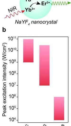
Schuck mentions another advantage of upconverting nanoparticles – namely, they operate by absorbing two or more infrared photons and emitting higher-energy visible light. "Since nearly all other materials do not upconvert, when imaging the UCNPs in a sample, there is almost no other autofluorescent background originating from the sample. This results into good imaging contrast and large signal-to-background levels." In addition, while organic dyes and Qdots can also absorb IR light and emit higher-energy light via a nonlinear two+ photon absorption process, the excitation powers needed to generate measurable two-photon fluorescence signals in dyes and small Qdots is many orders of magnitude higher than is needed for generating upconverted luminescence from UCNPs. "These high powers are generally bad for samples and a big concern in bioimaging communities" Schuck emphasizes, "where they can lead to damage and cell death."
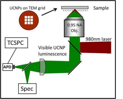
Schuck notes that two other key aspects central to the discoveries mentioned in the paper – using advanced single-particle characterization, and theoretical modeling – were a consequence of the multidisciplinary collaborative environment at the Foundry. "This study required us to combine single-molecule photophysics, the ability to synthesize ultrasmall upconverting nanocrystals of almost any composition, and the advanced modeling and simulation of UCNP optical properties," he says. "Accurately simulating and modeling the photophysical behavior of these materials is challenging due to the large number of energy levels in these materials that all interact in complex ways, and Emory Chan has developed a unique model that objectively accounts for all of the over 10,000 manifold-to-manifold transitions in the allowed energy range."
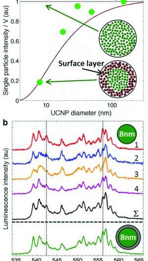
Previously, Schuck says that the conventional wisdom for designing bright UCNPs had been to use a relatively small concentration of emitter ions in the nanoparticles, since too many emitters will lead to lower brightness due to self-quenching effects once the UCNP emitter concentration exceeds ~1%. "This turns out to be true if you want to make particles that are bright under ensemble imaging conditions – that is, where a relatively low excitation power is used – since you have many particles signaling collectively," Schuck explains. "However, this breaks down under single-molecule imaging conditions." In their paper, the researchers have demonstrated that under the higher excitation powers used for imaging single particles, the relevant energy levels become more saturated and self-quenching is reduced. "Therefore," Schuck continues, "you want to include in your UCNPs as high a concentration of emitter ions as possible." This results in the nanoparticles being almost non-luminescent at low-excitation-power ensemble conditions due to significant self-quenching, but ultra-bright under single-molecule imaging conditions.

Another important implication of this finding, Schuck adds, is that it should change how people will screen for the best single-molecule luminescent probes in the future. "Until now," he notes, "people would first look to see which probes were bright using ensemble-level conditions, then would investigate only that subset as possible single-molecule probes. Our new probes would, of course, have failed that screening test!"
Schuck again emphasizes that "a key reason this discovery happened is that we have experts in all key areas in the same building, and we were able to quickly iterate through the theory-synthesis-characterization cycle."
Regarding future research directions, notes Schuck, the scientists are pursuing a few different avenues. "We'd certainly like to now use these newly-designed UCNPs for bioimaging....so far, we've only investigated the fundamental photophysical properties of these particles when they're isolated on glass. We believe one exciting and important application will be their use in brain imaging – particularly for deep-tissue in vivo optical imaging of neurons and brain function.
In closing, Schuck mentions other areas of research that might benefit from their study. "I think a primary application is in single-particle tracking within cells. For example," he illustrates, "labeling specific proteins with individual UCNPs and tracking them to understand their cellular kinetics."
Along different lines, Schuck adds, it turns out that UCNPs are also excellent probes of very local electromagnetic fields. "This is because lanthanides have a rather unique set of photophysical properties such as relatively prevalent magnetic dipole emission, allowing us to probe optical magnetic fields, and very long lifetimes such that transitions are not strongly allowed, which allows us to more-easily probe cavity quantum optical effects such as the Purcell enhancement of emission. In fact, Schuck concludes, an experiment that uses UCNPs to report on the near-field strengths and field distributions surrounding nanoplasmonic devices is just underway."
More information: Engineering bright sub-10-nm upconverting nanocrystals for single-molecule imaging, Nature Nanotechnology 9, 300–305 (2014), doi:10.1038/nnano.2014.29
Journal information: Nature Nanotechnology
© 2014 Phys.org


