Scientists rely on high-tech eyes to spy on microscopic world

It's been said that a picture is worth a thousand words, and at the Agricultural Research Service's Electron and Confocal Microscopy Unit (ECMU) in Beltsville, Maryland, this adage couldn't be more true. Led by unit director Gary Bauchan, the ECMU is tasked with producing high-resolution images that provide a window to the extraordinary world of the unseen.
"We have observed viruses, bacteria, fungi, nematodes, insects, mites, and parasites that threaten global food security, and we've contributed to the discovery of how pathogens spread by helping elucidate their relationship to the environment, hosts, and vectors," says Bauchan. "We've also described new biocontrol agents for the management of pathogens and characterized healthy and infected plant and animal tissues to discern the structural changes caused by pathogens."
Other ECMU capabilities include tracking the development of genetically transformed plants using fluorescently tagged plant cells and tissues and contributing to improved food safety by determining the mechanisms by which bacteria, fungi, and parasites infect fresh produce.
Scientists conducting multifaceted studies at ARS's Beltsville Agricultural Research Center (BARC) routinely call upon the microscopy experts in the unit to image all manner of specimens and samples. The researchers benefit not only in furthering their research objectives, but also by obtaining images to illustrate their journal articles, journal covers, grant proposals, web pages, and poster presentations.

Needless to say, the microscopy unit's services are in high demand and greatly appreciated. The latter was apparent from the turnout of BARC scientists and others who attended an October 11, 2012, ribbon-cutting ceremony presided over by Bauchan and Beltsville Area Director Joseph Spence. The event marked the completion of 2 years of renovations to the climate-controlled building that houses the ECMU and its prized collection of multi-million-dollar microscopes. Those include a low-temperature scanning electron microscope (LTSEM), a variable-pressure scanning electron microscope, two transmission electron microscopes, a confocal laser-scanning microscope (CLSM), and a Hirox digital video microscope. All are equipped with state-of-the-art digital cameras to speed the delivery of resulting images to researchers.
The ribbon-cutting ceremony also was in recognition of the expertise, creativity, and resourcefulness with which Bauchan, ECMU support scientists Charlie Murphy and Margaret Dienelt, and information technology specialist Christopher Pooley produce the images (at magnifications of up to 300,000 times) and ensure the integrity of the specimens for analysis by the researchers who submit them.
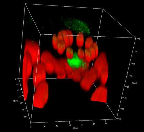
For example, when John Hammond, a plant pathologist at the U.S. National Arboretum's Floral and Nursery Plants Research Unit, contacted the team for assistance, the result was a three-dimensional image of how viruses spread from leaf veins to adjacent leaf cells. The image, which was generated using fluorescently tagged viruses and a Zeiss 710 CLSM, accompanied an article in the August 2012 issue of the Journal of General Virology and illustrated the cover of the November 2012 issue.
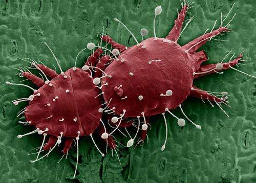
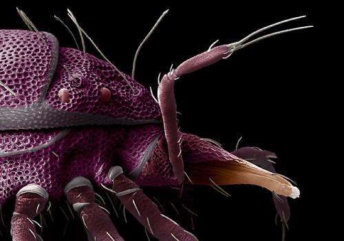
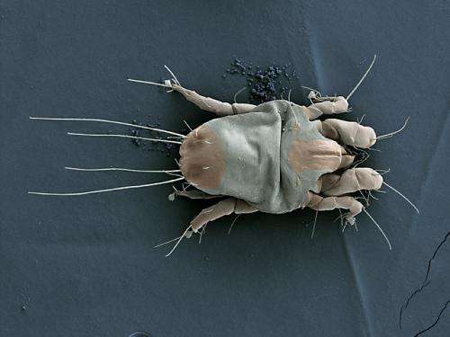
Each of the unit's microscopes offers unique capabilities and requires special handling to prepare specimens and samples prior to imaging. For systematic studies of mites, an LTSEM can be used. This necessitates flash-freezing a mite specimen (while still alive) at -321°F and coating it with platinum. In essence, the procedure freeze-frames the mite in time, allowing detailed examination of its features, physiology, behavior, and interaction with its immediate surroundings.
Imparting color to the high-magnification images helps further reveal critical, but often subtle, morphological differences between mite species, such as the size, shape, or number of setae (sensory organs) on their bodies. This capability has already proven invaluable to Beltsville scientists working with visiting scientist Jenny Beard, from the Queensland Museum in Australia, to differentiate species of mites in the genus Raoiella, members of which pose a significant threat to crops worldwide, including species of palm in the United States.

"At the Systematic Entomology Laboratory [SEL], our mite identifications support APHIS [USDA's Animal and Plant Health Inspection Service] plant protection and quarantine efforts, state agricultural agents, extension agents, and other countries looking for information and help on mite pests that affect their crops—or crops they export to our country," says mite expert Ron Ochoa, an ARS entomologist.
Images developed by SEL and ECMU have been used to develop an online identification key for flat mites of the world, tinyurl.com/flatmites. One year after the March 2012 launch of this website, there had been more than 86,392 visits to the web page, with inquiries from 196 countries. Ochoa notes that having access to the images is especially critical in making identifications that can become the impetus for regulatory decisions aimed at safeguarding U.S. agriculture, such as whether to treat, quarantine, or reject an agricultural import.

Bauchan estimates that the ECMU collaborated on 40 different projects last year, and 2013 looks to be just as busy—not only in literally answering the call of science, but also capturing it visually, as the sampling of images illustrating this article can attest.
More information: "Scientific Works of Art Reveal a Hidden World" was published in the August 2013 issue of Agricultural Research magazine. www.ars.usda.gov/is/AR/archive/aug13/
Journal information: Agricultural Research
Provided by Agricultural Research Service


